"The Flap Never Heals"
Below are three photos of an eye with a partially detached LASIK flap. The patient had LASIK in 2002. Several years later this eye developed corneal ectasia, requiring the patient to wear a hard contact lens. Due to a minor contact lens-related trauma which would have been uneventful in a normal eye, the LASIK flap separated from the underlying cornea in the 4:00 to 6:00 quadrant. The greenish-yellow dye which was instilled in the eye has penetrated under the flap. This case demonstrates the fragile state of the post-LASIK cornea, and proves that the flap never heals. (Click on top image to enlarge).


Below are three photos of an eye with a partially detached LASIK flap. The patient had LASIK in 2002. Several years later this eye developed corneal ectasia, requiring the patient to wear a hard contact lens. Due to a minor contact lens-related trauma which would have been uneventful in a normal eye, the LASIK flap separated from the underlying cornea in the 4:00 to 6:00 quadrant. The greenish-yellow dye which was instilled in the eye has penetrated under the flap. This case demonstrates the fragile state of the post-LASIK cornea, and proves that the flap never heals. (Click on top image to enlarge).

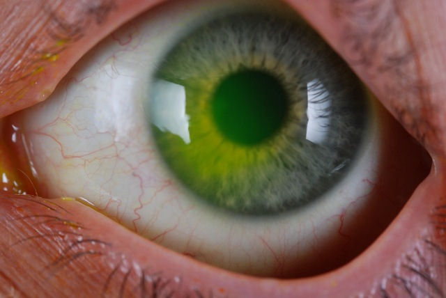
Below are two photos of an eye with a detached LASIK flap. The flap detached during contact lens fitting -- another example of how fragile the cornea is after LASIK surgery. In the first image, you can clearly see the dye which penetrated the space between the flap and the underlying cornea. LASIK patients are at life-long risk of flap dislocation, infection, epithelial ingrowth, DLK, and other complications. (Click on the photo to view a larger image.) In the second image, the red arrow points to a dark area of space where the flap is not attached to the cornea. This space runs along the flap margin.

 The LASIK flap margin is clearly visible in the photographs below. This is evidence that the flap never heals.
 Image below: Patient had LASIK in 2003.
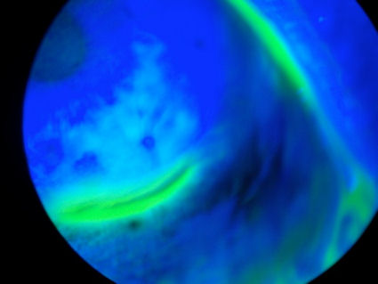
 The photos below were taken through a slit lamp (bio-microscope) after instillation of fluorescein dye. A blue light is used to illuminate the dye, which stains damaged areas of the cornea. The green-yellow circle around the periphery of the cornea is the LASIK flap margin. It is staining because the interface is still open, demonstrating the cornea's inability to fully heal.

 This next photo is the cornea of a patient who had LASIK in 2005. The deep staining at the margin of the LASIK flap indicates that there is an open portal at the cornea-flap interface, exposing the patient to infection from opportunistic organisms such as viruses and bacteria.
 The following images were acquired using a technique called optical coherence tomography, or OCT. The instrument produces a cross-sectional image by scanning the front of the eye (anterior segment) with a beam of light. Think of it like an ultrasound using light instead of sound waves to create an image of living tissues.
Below is a cross sectional scan of a cornea with a LASIK flap which has separated from the underlying cornea. The red arrow points to the location where the flap separated. The patient had RK followed by LASIK, and later developed ectasia. The patient is hoping to achieve functional vision with a specialty contact lens and avoid the need for corneal transplant.
 The cross-sectional scan below shows a cornea with post-LASIK ectasia and the flap separating from the underlying tissue. The green arrow points to the location of the ectasia, and the red arrow points to the location of the flap separation.

Below is a cross sectional scan of a cornea with a LASIK flap which has separated from the underlying cornea. The red arrow points to the location where the flap separated. The patient had RK followed by LASIK, and later developed ectasia. The patient is hoping to achieve functional vision with a specialty contact lens and avoid the need for corneal transplant.
Copied by:-http://www.lasikcomplications.com


